We all have heard about neurodegenerative disorders like Alzheimer’s, Parkinsons, ALS, or Multiple Sclerosis. But what we haven’t heard about is that the detection of these disorders demands a variegated approach on distinct layers to expose their complexities. This is where neuroradiology research and clinical trial consulting come into the big picture. Professionals leverage neuroradiology to spot the cause of neurological disorders, monitor disease progression, and more. However, how exactly does neuroradiology work, and how is it reshaping the way we understand everything from simple headaches to complex conditions like traumatic brain injury and strokes? Well, read on to find out!
Table of Contents
ToggleWhat Exactly is Neuroradiology?
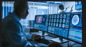
In layman’s terms, neuroradiology is a specialized field of radiology that focuses on diagnosing and treating conditions in the brain, spine, head, and neck using different imaging techniques, such as MRIs and CT scans. With these images, professionals can get a closer look at what’s happening inside your head and spot problems that would otherwise be invisible.
So, the human brain is super complicated. In fact, it’s like the control center for everything in your body, from movements to emotions and memories. Before the rise of neuroradiology, the only way to study the brain’s structure was through surgery or, in unfortunate cases, autopsy, which was way too risky and limited in its applications.
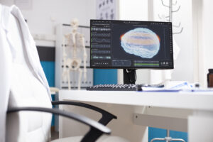
However, today, with advanced imaging techniques like MRI (Magnetic Resonance Imaging) and CT (Computed Tomography) scans, neuroradiology allows us to see the brain in astonishing detail, examine its soft tissues, such as white and gray matter, and identify abnormalities such as tumors, lesions, or areas affected by strokes for better patient outcomes.
Mapping Brain Functions
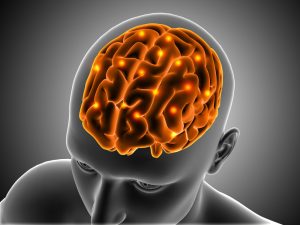
One of the most exciting aspects of neuroradiology is helping us understand how the brain functions with unbiased and accurate outcomes. For instance, functional MRI or fMRI is quite a game-changer. Unlike conventional MRIs, which show brain structure, fMRIs highlight brain activity. When certain parts of the brain “light up” during specific tasks, like speaking, thinking, or moving, the fMRI captures that.
Researchers can use the data from fMRI to map out the regions of the brain responsible for particular functions. Thanks to neuroradiology and fMRI, we now know which areas of the brain handle language, emotions, and decision-making. This might sound trivial on paper, but when it comes to brain surgeries, it allows surgeons to avoid critical areas, and even this data can be used in medical malfunction cases where a surgeon did not consider the neuroradiological aspects of a patient before operating.
Breaking New Ground in Mental Health
Mental health conditions, like depression and anxiety, have often been difficult to understand because we couldn’t see what was happening inside the brain. Neuroradiology is changing that. By using brain scans, researchers are now able to see the structural and functional differences in the brains of people with mental health disorders. For instance, brain scans have shown that people with depression may have reduced activity in certain areas, helping doctors create more personalized treatment plans.
Understanding Neurodegenerative Diseases
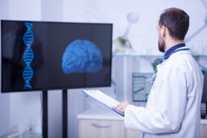
Neuroradiology is also giving us critical insights into neurodegenerative diseases like Alzheimer’s and Parkinson’s. Doctors can now detect changes in the brain’s structure long before symptoms appear. This early detection can lead to better treatments and, hopefully, one day, prevention.
Looking for help with neuroradiology research and clinical trial consulting? Reach out to the team at Neuro Experts today and get started!