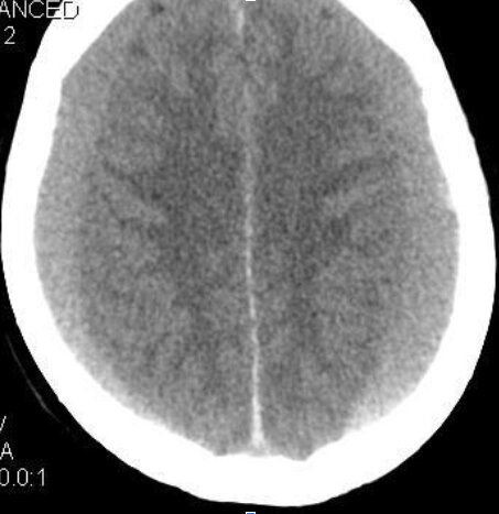
A 78-year-old male with history of multiple prior falls is brought into the local ED from his care facility because his caretaker noticed his altered mental status. A head CT was obtained.
Table of Contents
Toggle
What type of head injury is seen in this image, and what space does this injury involve?
- Epidural, extra-axial
- Epidural, intra-axial
- Subdural, extra-axial
- Subdural, intra-axial
- Subarachnoid, extra-axial
- Subarachnoid, intra-axial
Subdural hematoma
- Non-contrast CT is the best initial study. MR is better able to depict extent and age.
- CT findings: Extra-axial collection of blood located between inner border cell layer of dura and arachnoid with a crescentic shape that spreads diffusely across cranial convexities as it can extend across cranial sutures but not dural attachments.
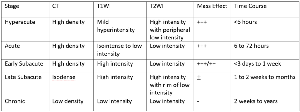
- Hyperacute phase (<6 hrs) can be of heterogeneous density or hypodense on CT
- Acute phase (6h – 3d) hematoma is homogeneously hyperdense ~60% of the time and mixed hyper- and hypodense 40% of the time.

Taken from Neuroradiology: The Requisites 3rd Edition (2010). Rohini Nadgir, David Yousem, Robert Zimmerman, Robert Grossman,

Case courtesy of A.Prof Frank Gaillard, Radiopaedia.org, rID: 36065
- MR findings: Variable in appearance secondary to the different appearance of blood on MR depending on its age.
- Typically, subdural hemorrhage is secondary to traumatic tearing of bridging cerebral veins. The trauma may be very minor, especially in elderly patients as they are predisposed tearing secondary to cerebral atrophy.
- Patient’s with ventricular shunts are at higher risk due to the shunted system not acting as a natural tamponade.
- Can grow slowly with increasing risk for mass effect and herniation if not identified and treated early.
- Recurrent hemorrhage is common.
- SDH is a neurological emergency that often requires surgical treatment, however the decision between operative or non-operative management is based on multiple factors. Typically, If size is < 1cm with no cerebral edema, they can be managed non-operatively.
- High-dose mannitol is often given to decrease risk of brain herniation prior to surgery.


